Home

Photoelectric Doctrine Prophet vitreous veils weather mental together

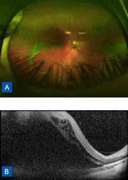
Retinal Detachment Asheville | Posterior Vitreous Detachments

Vitreous Veils with Sickle Cell Trait Disease | Eccles Health Sciences  Library | J. Willard Marriott Digital Library
Vitreous Veils with Sickle Cell Trait Disease | Eccles Health Sciences Library | J. Willard Marriott Digital Library

Full article: Intermediate Uveitis: A Review

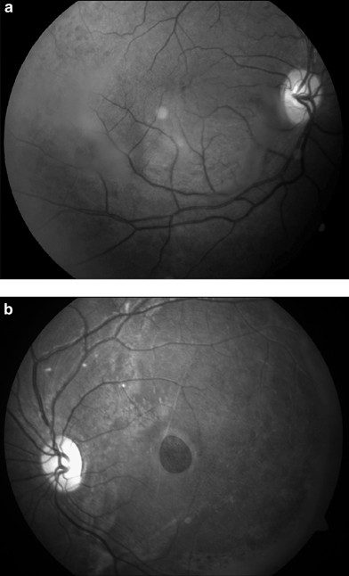
Macular hole secondary to X-linked retinoschisis | Eye

X-Linked Retinoschisis without Macular Retinoschisis: A New RS1 Mutation -  Ophthalmology Retina
X-Linked Retinoschisis without Macular Retinoschisis: A New RS1 Mutation - Ophthalmology Retina

Treatment of Floaters - Scott E. Pautler, M.D. TampaScott E. Pautler, M.D.  Tampa
Treatment of Floaters - Scott E. Pautler, M.D. TampaScott E. Pautler, M.D. Tampa

Atlas of Ophthalmology

María Iglesias on X: "Amazing vitreous veil in the june #eye cover  #ophthalmology https://t.co/g9R7UqoU9w" / X
María Iglesias on X: "Amazing vitreous veil in the june #eye cover #ophthalmology https://t.co/g9R7UqoU9w" / X

Blurry Vision in a Young Patient | Duke Health Referring Physicians

Atlas Entry - Anterior vitreous inflammation in autoimmune retinopathy

Heredofamilial Vitreoretinopathies - RETINA AND VITREOUS - Albert &  Jakobiec's Principles & Practice of Ophthalmology, 3rd Edition
Heredofamilial Vitreoretinopathies - RETINA AND VITREOUS - Albert & Jakobiec's Principles & Practice of Ophthalmology, 3rd Edition

Discover images - Retina Image Bank

Color fundus photographs of Case 1. The right eye (A) demonstrates a... |  Download Scientific Diagram
Color fundus photographs of Case 1. The right eye (A) demonstrates a... | Download Scientific Diagram

Case 2: Color fundus photographs of the left and right eyes showing... |  Download Scientific Diagram
Case 2: Color fundus photographs of the left and right eyes showing... | Download Scientific Diagram

Clinical features of Goldmann–Favre vitreoretinal degeneration -  ScienceDirect
Clinical features of Goldmann–Favre vitreoretinal degeneration - ScienceDirect

X-linked Retinoschisis - EyeWiki

Juvenile X-Linked Retinoschisis | Ento Key

Vitreous I Flashcards | Quizlet

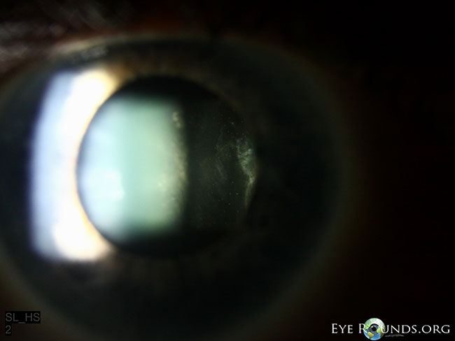
Congenital Vitreous Veil

Ophthalmology-Notes And Synopses - Fundus photograph shows inferiorly  located snow ball opacities, vitreous veils and vascular sheathing in a  patient with pars planitis. | Facebook
Ophthalmology-Notes And Synopses - Fundus photograph shows inferiorly located snow ball opacities, vitreous veils and vascular sheathing in a patient with pars planitis. | Facebook

Moran CORE | Stickler Syndrome

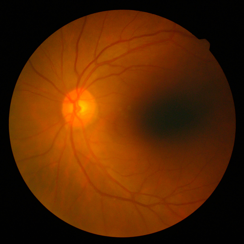
Moran CORE | Stickler Syndrome

Moran CORE | Stickler Syndrome

Diseases of the Retina and Vitreous | Ento Key

Clinical Features and Course of Ocular Toxocariasis in Adults | PLOS  Neglected Tropical Diseases
Clinical Features and Course of Ocular Toxocariasis in Adults | PLOS Neglected Tropical Diseases

Fundus image showing submacular exudation RE (arrow mark) and foveal... |  Download Scientific Diagram
Fundus image showing submacular exudation RE (arrow mark) and foveal... | Download Scientific Diagram

X-linked Retinoschisis - EyeWiki